Crush your head
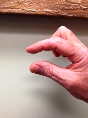
I often find that patients and even physicians have widely differing views on the size of blood clot when they get an ultrasound report or interpretation. There are several types of blood clots and they can occur in both arteries and veins. Today, we are talking about blood clots in the veins of the lower extremity or legs. One of the most common questions I ask patients is “how big do you think a blood clot is”? A common response is for the patient to hold up their thumb and index finger as if they were measuring about an inch (crush your head).
While clots can be all sizes and can range from a few mm. to 80+ cm. it is not uncommon for a clot or thrombus to extend the length of the vein that it is in. Another common statement I hear from patients is “I have five blood clots in my leg”. In actuality they usually have five veins that have thrombus or blood clots. They could have one clot that extends into five veins or the clots could be isolated in several veins and not all connect. I don’t like it when a physician tells a patient that they have 2, 3 or 5 clots. This terminology is meant help the patient understand, however often complicates the situation. When properly explained patients get a better understanding of the situation. With venous disease there are various patterns in regard to the progression of thrombus, however it is most common for the thrombus to start in the calf and extend up the leg or start in the pelvis and extend distally (less common of the two).

There is wide variation among providers and specialties in how to treat blood clots below the knee. I have found that it is important to describe to the referring physician the extent in which the blood clot is found and not just the vein by name. Here are two examples of how a venous duplex report may be interpreted. “Acute deep vein thrombus identified in the posterior tibial vein(s)“. In this case the referring physician has little understanding of the extent or location, except that it is in the calf. On the other hand if the report stated the findings like this: “Acute deep vein thrombus identified in both posterior tibial veins, the thrombus is occlusive and extends from the ankle to the tibial/peroneal trunk in the proximal calf”, the provider would have a clear understanding that the thrombus (is not the size of an inch) and has extended the entire length of the calf. Many clinicians or organizations discount the dangers of below the knee thrombus, however by giving them more detailed information they can make a more informed decision. This type of detail also helps if the patient is still symptomatic at a later time and has another ultrasound to compare.

These larger blood clots often when not diagnosed are fatal. I have had a patient walk in the door with a blood clot extending from the calf all the way to the mid abdomen (IVC). Recently a California man had a 24 in. or 60 cm. clot removed via a device called an AngioVac. The thrombus extended from his legs into the heart. There are several such devices on the market; these types of advancements have increased the effectiveness of treatment for larger
blood clots. For blood clots or DVT that are not obstructive in the thigh or pelvis the use of anticoagulation drugs (blood thinners) is common. We will talk about the various kinds of blood thinners available in another blog.
Categories: Banter, diseases, health, medical imaging, phlebology, radiology, ultrasound education, vascular surgery, vein disease, women's health